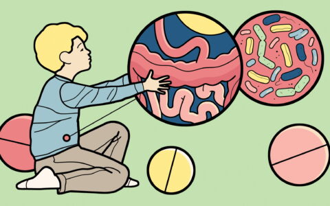
Eosinophilic esophagitis (EoE), which can lead to fibrostenotic strictures and dysphagia, is estimated to affect around 57 per 100,000 people in the United States, and rates are growing.
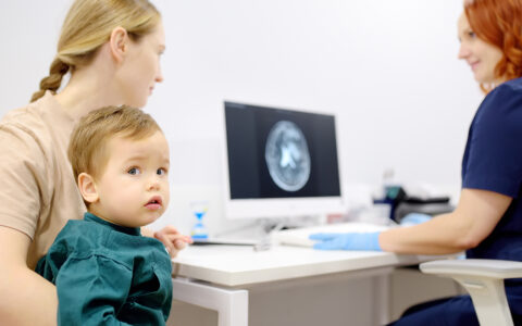
Assessment of the esophageal lamina propria — the loose connective tissue layer that lies beneath the epithelium — is an important means of tracking EoE complications. Although mucosal biopsies are routine for these patients, deeper structures such as the epithelium are sampled in only about half of the cases due to associated risks of tissue damage.
“Our tool predicts probability of lamina propria fibrosis with 84 percent confidence.”
Now, a web-based tool is available that reliably predicts the probability of lamina propria fibrosis when samples are unavailable for histopathology. This tool was developed by Girish Hiremath, M.D., a pediatric gastroenterologist at Vanderbilt University Medical Center, and colleagues in the Consortium of Eosinophilic Gastrointestinal Disease Researchers.
Their Model to Predict Lamina Propria Fibrosis in Eosinophilic Esophagitis uses age and three esophageal epithelial features.
“Our tool predicts probability of lamina propria fibrosis with 84 percent confidence,” said Hiremath, who was first author on a validation study published in the American Journal of Gastroenterology. “With that level of certainty, clinicians can make more informed choices about treatment and risk-stratify their patients for future fibrostenotic complications.”
Battling the Allergens
In EoE, an allergen enters the esophagus, prompting an excessive influx of eosinophils. These cells can release large quantities of toxins, leading to uncontrolled and chronic inflammation.
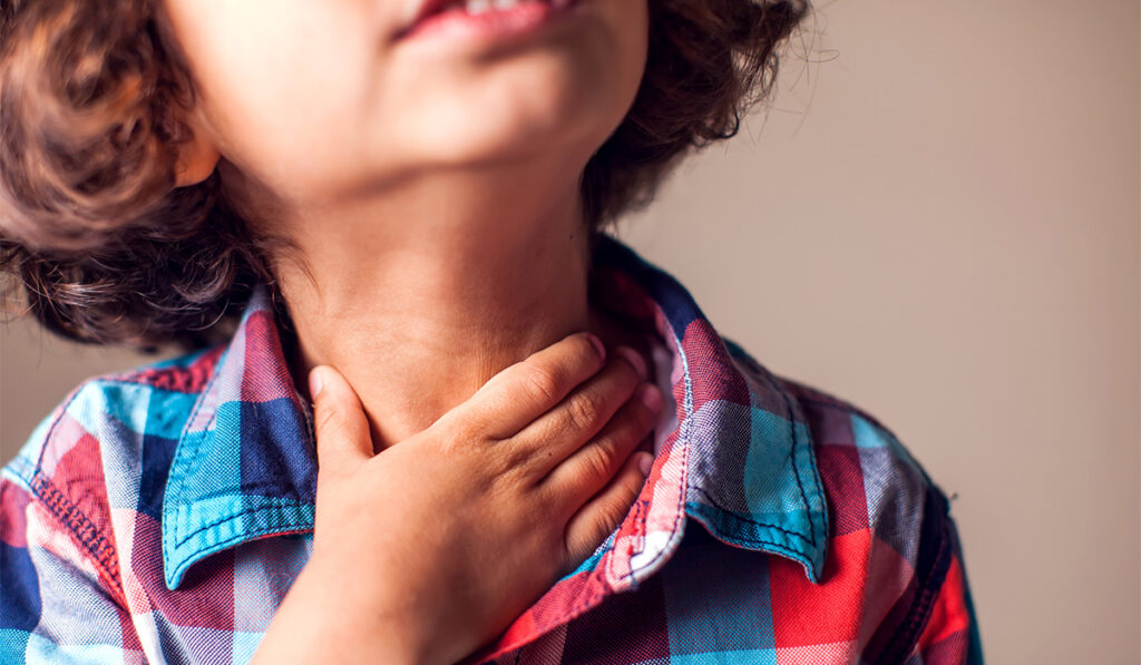
The disease manifests as early as in infancy and is more prevalent in males. Because it shares some symptoms with gastroesophageal reflux disease, often it is not diagnosed or treated early. Even when this happens, fibrotic changes in the lamina propria may develop, potentially leading to food impaction, strictures, narrowing of the esophagus and difficulty swallowing.
Current medical treatments, which include swallowed steroids and injectable biologics, are not curative but help combat the inflammatory effects of allergens, control symptoms and slow progression. Some patients can remit symptoms through dietary elimination of foods suspected of triggering esophageal inflammation.
In severe cases, endoscopic procedures to mechanically dilate the esophagus are necessary to provide some relief for patients. Hiremath says people with EoE require frequent endoscopies to diagnosis and monitor the disease, and some have up to four procedures per year.
Study and Confirmation
To develop and validate their new webtool, the researchers obtained data from 419 patients ages three years or older, examining a total of 1,253 biopsies taken at all stages of EoE.
“Previously, we had found a high concordance between the presence of surface epithelial alteration and dyskeratotic epithelial cells and lamina propria fibrosis in children with EoE,” Hiremath said. “For this study, we then hypothesized that the degree of epithelial feature abnormality can predict fibrosis in biopsies where it is impossible to determine the status of lamina propria.”
Originally, the team looked at the seven histologic variables present on the pathology reports, but ultimately narrowed it down to four metrics: age, surface epithelial alteration, dyskeratotic epithelial cells and basal zone hyperplasia.
In the 284 patients whose biopsies included lamina propria, researchers looked at the histologic findings of fibrosis. First, they tested the model on those with fibrotic changes, per the four-variable scale. They then applied the model to the patients whose lamina propria biopsies had no histologic signs of fibrosis.
“We found an AUC of .84,” Hiremath said. “That was tremendous.”
The team confirmed the validity of the tool using data comprising demographic, clinical, and histologic information of children (aged 3–18 years) with EoE undergoing biopsies via esophagogastroduodenoscopy at Monroe Carell Jr. Children’s Hospital at Vanderbilt.
The researchers then put their tool on the web so it is usable by practitioners all over the world. To determine probability of fibrosis, the clinician inputs data for the four metrics graded on a 0-3 scale, entering them separately by grade and stage. The tool delivers a probability based on each. The model assumes use of the Eosinophilic Esophagitis Histology Scoring System to assess severity and extent of diseased tissue.
Future Prediction
“The predictor epithelial features were in strong agreement across the biopsy sites, and the performance of our model was not confounded by treatment status or exposure to swallowed topical steroids,” Hiremath said.
“The performance of our model was not confounded by treatment status or exposure to swallowed topical steroids.”
Hiremath says that the cross-sectional data they analyzed suggests that the fibrostenotic complications can occur in EoE in a trackable, time-dependent manner.
“However, using or developing the tool so it predicts future changes will require more studies, ” he said.