In the context of oncologic surgery, it can be difficult to provide a detailed pathological assessment of a resected specimen to a surgical team waiting in the operating room.
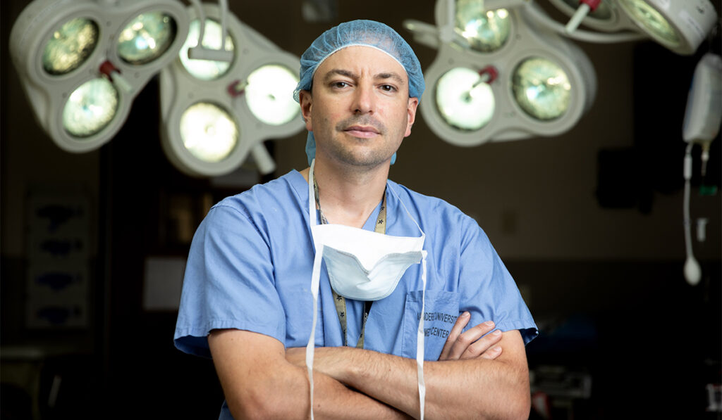
This difficulty prompted head and neck surgeon Michael Topf, M.D., M.S.C.I. and his team at Vanderbilt University Medical Center to devise a way to capture important details of specimens for quick transmission to the waiting surgeons.
“I was inspired based on an unmet clinical need,” Topf said. “There was an opportunity for improvement in communication among the multidisciplinary cancer team.”
Topf says the need is particularly great for head-and-neck cancer surgeries that involve complex tissues about which surgery and pathology teams cannot always communicate in person.
Topf and his team developed and tested a 3D scanning and analysis technique, incorporating it into the surgical pathology workflow. The feasibility of their approach is described in a prospective study reported in the journal Head & Neck.
This technique not only enables the preservation of visual details of surgical specimens, which often become destroyed during processing, but it also could enhance the information exchange between surgeon and pathologist regarding important details, the authors said.
Real-time Specimen Mapping
The team aimed for a protocol that was quick to perform with minimal impact on the pathology lab’s workflow. In a key step, surgical specimens will undergo 3D scanning to obtain surface topography data prior to further processing. Computer-aided design software provides an annotated map of the specimen, showing the sites of sectioning, inking, and margin sampling that can be shared with the surgical team through teleconferencing.
In their study, Topf and his team used the approach with surgical samples from 40 patients with head and neck neoplasms. The team evaluated outcomes related to the protocol’s practicality and surveyed two dozen surgical and pathology personnel participating in the process to get their feedback.
Feasibility and Impacts
Topf and his colleagues obtained intraoperative 3D scans and developed 3D models for all fresh surgical specimens in the study, with image acquisition adding about eight minutes to the sample processing timeline.
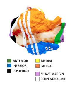
The data suggested the team’s approach aided communication between the pathology and surgery teams about details, such as the margins in tumor resection, while also preserving the image for potential review at a later time.
Topf said the process has been used over 200 times. Since publishing their study, the team has expanded to involve other cancer types and made other refinements.
“This protocol is transferable to any solid malignancy that surgery plays a role in,” Topf said.
A Team Approach
Traditionally, most members of a care team have no contact with or visual image when making decisions about a resected specimen.
“This protocol empowers other people on the team to learn about the patient’s cancer.”
“This protocol and the work that we are doing really empowers other people on the team to learn about the patient’s cancer and allows us to communicate better and make advancements to improve patient care,” he explained.
Reflecting on other recent observations from the team, Topf said it surprised him how often these specimen maps provide crucial clarity on the site of margin sampling.
“That means in cases where we don’t use it, we could be getting it wrong.”
Visualizing Surgery’s Future
Topf said he looks forward to studying impacts of these changes on patient outcomes. He and his colleagues are investigating use of an augmented-reality headset to allow an expert pathologist to virtually enter the operating room through teleconferencing.
“As the hardware and the software continues to improve, we can envision that surgery in the future could be fundamentally changed.”
Additionally, Topf, in collaboration with Jie Ying Wu, Ph.D., an assistant professor of engineering at Vanderbilt, and other colleagues, has been exploring the use of augmented reality as a visual aid in surgeries. In the Annals of Surgical Oncology, the team recently described an augmented-reality headset that could superimpose 3D specimen holograms over surgical sites to assist with re-resection.
“I don’t think it’s quite ready for routine clinical use yet, but as the hardware and the software continues to improve, we can envision that surgery in the future could be fundamentally changed,” Topf said.