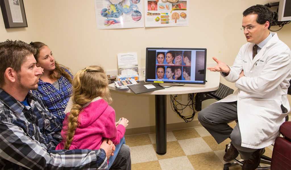
Scott Stephan, M.D., a facial and plastic reconstructive surgeon at Vanderbilt University Medical Center, is pioneering a new technique for microtia reconstruction using 3D-bioprinted cartilage tissue.
Stephan, who directs the medical center’s Microtia and Atresia Clinic, says that with recent advances in cartilage-tissue engineering, using a patient’s own chondrocytes to provide customizable outer-ear repair may soon become a reality.
“The gold standard for microtia has been surgical repair using costal cartilage and porous polyethylene construct, but the paradigm is shifting,” Stephan said. “The latest advances in 3D bioprinting technology have laid the groundwork for achieving native cartilage architecture and function.”
The ‘Auregen’ Story
In recent years, 3D bioprinting has emerged as a promising new approach for producing complex biological constructs. In 2016, Stephan was approached by a biotech startup, Auregen Biotherapeutics, to help solve a difficult problem — engineering auricular cartilage tissue using a patient’s own chondrocytes.
After several months, Stephan and the Auregen team came up with a possible solution.
“By combining a biodegradable polymer scaffold with a tiny piece of the patient’s cartilage tissue, we’re able to grow native auricular cartilage in the lab,” Stephan said.
According to Stephan, 3D-bioprinted cartilage offers the potential for case-by-case customization, which is particularly useful during operative planning, and can help surgeons better understand each patient’s unique anatomy, thereby improving aesthetic outcomes.
“Patients often have unique anatomical abnormalities that can preclude favorable aesthetic results following reconstructive surgery,” he explained.
Moving to New Standard
Currently, microtia repair techniques can be divided into two major categories — autologous and synthetic reconstruction.
Stephan notes that both techniques are highly complex and require specialized expertise and that a controversy exists as to which approach is superior.
A less complicated surgical procedure may be one advantage of a 3D-bioprinted, custom implant, Stephan adds. Abnormalities corrected by traditional techniques have included mandibular hypoplasia, a deficient mastoid and asymmetric malar height, and projection, among others.
“Serving patients is an extremely gratifying yet humbling undertaking, as each case comes with its own unique set of challenges,” he said.
As the team continues to develop its proprietary 3D tissue-engineered method, Stephan believes this approach could become a new standard of care, although additional work remains.
Building Bridges in Research
For next steps, Auregen wants to build international bridges in research, aiming to partner with more experts to help bring this technology to the clinic, Stephan says.
“To achieve optimal outcomes, centers of excellence and those that manage a higher volume of microtia cases are important for this specialized surgical service,” Stephan said.
Auregen is also seeking to gain full FDA approval of its implant technology, and clinical trials are underway, including in pediatric patients.