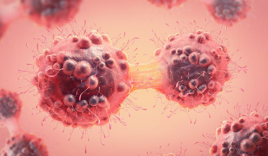
Mutations in SETD2, an important factor in DNA transcription, have been directly implicated in cancer in recent years, based on tumor cell sequencing and the cancer genome atlas program.
Now, Frank Mason, Ph.D., an assistant professor of hematology and oncology, and Kimryn Rathmell, M.D., Ph.D., chair of the Department of Medicine at Vanderbilt University Medical Center, are connecting the dots between mutations expressed in the enzyme SETD2 and errors in chromosome organization. Their most recent work was published in the Proceedings of the National Academy of Sciences.
Using genetic and pharmacological approaches in human cervical cancer cells and mouse fibroblasts, the researchers associated SETD2 loss or inhibition with the formation of specific types of defective chromosomes, including isochromosomes that contain mirror-imaged inversions.
“These abnormal chromosome structures are known to be present in a number of cancers and developmental disorders,” Mason said. “But how these abnormal chromosomes, which causes whole chromosome arms to be lost, arise is unknown. SETD2 loss or mutation is the first link between a tumor suppressor and their formation.”
Further, they demonstrated for the first time, that disruption of SETD2 during DNA replication is the sequalae of a defective mitosis, leading to errors in chromosome segregation.
This discovery sets the stage for uncovering how SETD2 protects mitosis and how its loss leads to defective chromosome production.
“There are likely multiple parallel activities that enforce a normal mitosis. Understanding SETD2 function and loss in detail may lay out the mechanism for how other mutagenic actors function and uncover ways to interfere with the defective chromosome formation,” he said.
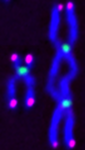
Three Chromosomal Defects
Three types of defective chromosomes have been associated with SETD2 loss, Mason explained: isochromosomes, dicentric chromosomes and acentric chromosomes.
While the mechanism for isochromosomes formation is unknown, the result is a chromosome that is a mirror image repeat. Dicentric chromosomes form when the strands have two centromeres instead of one. In this case, two microtubules are in a tug of war, stretching the chromosome, and damaging the DNA. Acentric chromosomes form when the strand has no centromere for a microtubule to attach to, so this chromosome fails to be incorporated into a daughter cell and is ultimately lost.
The team’s work shows that the ability for SETD2 to methylate histones, the spool-like structure around which the DNA strands wrap, is critical for preventing the formation of these disrupted chromosomes.
SETD2 Loss
Rathmell, who was recently named director of the National Cancer Institute, has been studying SETD2 for years, trying to uncover what it is about loss of SETD2 that promotes tumorigenesis in kidney cancer. For this study, Mason and she used an inducible CRISPR and conditional degron system to delete SETD2 at specific cell cycle stages, enabling them to see the immediate early defects, and then, in subsequent cell divisions, their consequences.
“SETD2 loss is involved in all these DNA damage responses, in all the stages.”
“When we deleted SETD2, we saw a host of problems in chromosome segregation,” Mason said. “And while we know SETD2 plays important roles during mitosis, we now know that a number of these issues in chromosomes segregation are the consequences of errors during DNA replication.”
SETD2 is also a mediator of the DNA damage repair response, regulating homologous recombination, which requires the recombinase RAD51. While previous groups identified that SETD2 loss disrupts RAD51 activity, Mason and his colleagues identified that RAD52, which mediates chromosomal mutations, takes its place.
“There are many ways that you can repair the DNA, depending on what stage of the cell cycle you’re in, and what type of damage it is. But SETD2 loss is involved in all these DNA damage responses, in all the stages,” Mason said.
“SETD2 may be recruited to these sites to perform DNA repair. More likely, though, SETD2 puts a methyl mark on the chromatin to recruit other repair factors to the site. If SETD2 is absent, alternative pathways that are more mutagenic appear to occur.”
Chemotherapy Resistance
Chromosomal instability is a major driver of chemotherapeutic resistance. Additionally, cells with the chromosomal aberrations are more resistant to chemotherapy and induce genetic heterogeneity, “similar to tumors that exhibit intratumoral phenotypic and genetic heterogeneity,” Mason said.
“In acute lymphoblastic leukemia, SETD2 mutations are more common after chemotherapy. This may be because chemotherapy is inducing mutations, but we suspect the mutated cells that were there before therapy are more resistant, so those are the ones that multiply,” Mason said.
Mason and Rathmell are trying to identify the pathways involved in promoting resistance once SETD2 is lost. The hope is to pinpoint therapeutic vulnerabilities in these mutated cells and interfere with the pathways that enable cancer cells to repair their mutant DNA, so that they die instead of multiplying.
Further Work in Stratification
Mason says that depending on the phase of the cell cycle, SETD2 might be doing different things, so the team is using a new degron system for acute protein degradation to look even more closely at each phase of the cell cycle in isolation.
“We still need to identify the mechanisms by which SETD2 protects DNA replication, and this could be the technique that allows us to unlock other mechanisms by which these isochromosomes and dicentric and acentric chromosomes form – ultimately, we hope, helping us find a way to prevent them from forming.”