Neuroimaging research by Francesca Bagnato, M.D., Ph.D. is poised to make possible in vivo assessment of neurodegeneration biomarkers for patients with multiple sclerosis (MS).
Bagnato, an associate professor of clinical neurology at Vanderbilt University Medical Center, is narrowing in on magnetic resonance imaging (MRI) biomarkers that are both sensitive and specific to myelin and axonal injury.
“If we push these MRI technologies, we’ll make the in vivo evaluation of neurodegeneration a reality.”
To find such markers, Bagnato is employing high-resolution quantitative MRI methods.
“If we push these MRI technologies, we’ll make the in vivo evaluation of neurodegeneration a reality, causing a turning point in the care of patients with MS and neurodegenerative disease,” Bagnato said.
Hunt for Biomarkers
Bagnato explained that the fundamental cause of irreversible disability in MS, neurodegeneration, comes secondary to the death of neurons and the breakdown of neuronal wires and their covers, called axons and myelin, respectively. The inability to detect neurodegeneration in patients makes it difficult to develop new treatments. For this reason, MS remains an incurable disease.
Research into MRI biomarkers of neurodegeneration is a top priority in the field, Bagnato says.
“Finding these biomarkers will set us on a path toward not only halting neurodegeneration but promoting neuroprotection in MS.”
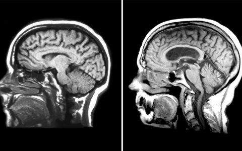
Among the MRI techniques her group is focused on for detecting and monitoring myelin and axonal injury is selective inversion recovery quantitative magnetization transfer imaging (SIR-qMT) and multi‐compartment diffusion MRI using the spherical mean technique (SMT).
As described in several reports published over the last five years, her team used these imaging approaches to evaluate patients with MS and healthy volunteers. They found their SIR-qMT and SMT protocols, used at 3-T, predict radiological and clinical disease severity.
“These types of images can be obtained using 3-T systems which are available at most institutions, making this screening strategy clinically feasible.”
Now, Bagnato and her team are applying these techniques to people recently diagnosed with MS to understand how early neurodegeneration occurs and how it may impact the likelihood of progression.
“Part of the excitement for these results is that these types of images can be obtained using 3-T systems which are available at most institutions, making this screening strategy clinically feasible,” Bagnato explained.
Toward Neuroprotection
After studies are completed, Bagnato plans to deliver size computations and MRI protocols for proof-of-concept clinical trials on neuroprotection and repair.
“We are building these imaging protocols to detect neurological dysfunction early, even before symptom onset, to open a wide window for altering disease progression in a timely manner, when there is still salvageable brain tissue,” Bagnato said.
In addition to clinical monitoring, she envisions these advanced MRI techniques as soon being used to better understand the fundamentals of MS progression, including modifiable and non-modifiable risk factors.
“In the very near future, I believe advanced MRI methods like those developed by our group and others will significantly improve our ability to study, monitor and treat MS,” Bagnato said. “That’s our vision. That’s our goal.”