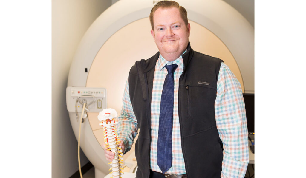
Seth Smith, Ph.D., associate director of the Vanderbilt University Institute of Imaging Science (VUIIS), was recently elected to the College of Fellows of the American Institute for Medical and Biological Engineering (AIMBE). The selection recognizes his work in advanced multiparametric MRI for translational applications, as well as his advocacy on behalf of first-generation scientists.
Discoveries spoke with Smith about his role in developing advanced MRI and his contributions to its evolution that hold promise for earlier and more definitive diagnosis of diseases of the spinal cord, optic nerves and other tissues.
Pioneering Contributions to MRI Imaging
Discoveries: Let’s talk first about your selection to the AIMBE College of Fellows, an entity only 2 percent of the professionals in your field are invited to join. What led to this recognition?
Smith: First of all, I am really honored to be selected, and I am a strong proponent of AIMBE’s mission to advocate with Congress and other lawmakers about the importance of imaging research in understanding disease evolution, treatment and cures.
MRI research is still a fairly young science, so I’ve had the opportunity to grow with the technology and do my part to push it forward. It has been largely focused on the brain, trying to understand its structure, function and abnormalities, but the rest of the body hasn’t received the same level of attention.
My contributions have focused on developing MRI research techniques, acquisitions and analysis for the spinal cord, but also other smaller neurological structures, such as the optic nerve. These and tiny nerves elsewhere in the body have been difficult to image by any modality.
Much of what I do involves developing advanced tools to look at the integrity of the spinal cord and at its relationship to neurological outcome, always with an eye towards the brass ring of clinical translation. For example, we need better means of diagnosing and understanding diseases like multiple sclerosis (MS), which significantly involves these small nerves and can take years to diagnose.
Discoveries: What first got you interested in small structure imaging?
Smith: I started my career working under Drs. Peter van Zijl and Hugo Moser at the Kennedy Krieger Institute, both of whom had an interest in developing spinal cord imaging for leukodystrophies. My interest was further sparked by the story of Lorenzo Odone (about whom the movie, Lorenzo’s Oil, was made), who had adrenoleukodystrophy (ALD).
During my Ph.D. training, Dr. Moser became intrigued by the fact that a variant of ALD called adrenomyeloneuropathy (AMN) affected the spinal cord but didn’t give the MRI signatures that would enable us to easily detect the disease. He asked me to try to develop something that would be useful for detecting AMN. So, I spent my Ph.D. developing tools to try to better image these tiny, difficult-to-image areas and diseases of the spinal cord.
Because of the successes we had with AMN, the lab started to collaborate with groups from the MS and neuro-ophthalmological communities, because the optic nerve and the spinal cord may hold information that is critical to understanding disease evolution that we aren’t able to see with conventional MRI. Upon moving to Vanderbilt, Dr. John Gore further supported these studies, and that’s where our research started to expand.
Focus on Sensitivity and Usability
Discoveries: Where have these last two decades taken you in this MRI evolution?
Smith: Our group and many others across the world have developed advanced MRI capabilities that provide information that far surpasses what conventional MRI can do. Advanced multiparametric imaging may offer insight into myelin or axonal health, as well as tissue microstructure through the diffusion of water, or functional activation through changes in oxy- versus deoxyhemoglobin. Since MRI is performed using pulse sequences, we have worked to tailor those sequences to be sensitive to different things in the spinal cord.
While we perform these developments at VUIIS, we work closely with vendors, as these methods aren’t yet a part of the standard vendor package for clinical MRI machines. I like the adage that for something to be clinically valuable it has to be useful, usable and used. The imaging that is being developed, while potentially useful and usable, is not yet used.
So, our research has two sides. One is to make the imaging sequences more sensitive to the specific pathologies of interest and the second is trying to improve speed and usability in a way that makes it clinically viable.
Avoiding “MS Mimickers” to Shorten the Diagnostic Odyssey
Discoveries: What gaps do these different imaging parameters fill in diagnosing MS and similar diseases?
Smith: With multiparametric imaging, we have the potential to examine both the spinal cord and the optic nerves for markers of myelin, axonal and vascular health that pertain to the development or evolution of MS. In fact, the Optic Neuritis Treatment Trial showed that if you see optic nerve damage and find a single brain lesion, the patient is almost 60 percent likely to develop MS.
Add to that the fact that the ophthalmology community has found that patients who have optic neuritis – even if they don’t have any sort of lesions in the brain – still have a one in four chance of developing MS at some point later in their life. Developing improved imaging tools to characterize these very small structures has become important for getting a jump on diseases like MS.
Discoveries: What is now on the horizon in your research?
Smith: One of the big wins for MS imaging right now is the identification of what is called central vein signs using susceptibility weighted MRI (SWI). It has been shown that a lot of MS lesions in the brain have a vein that runs through them. Thus, activity of MS lesions is coupled somehow with vascular compromise.
Assuming the spinal cord and brain are not that distinct and understanding that the spinal cord is also involved in MS, we are trying to find the same abnormal vasculature in spinal cord lesions. Using one of our specialized systems here – a 7-Tesla MRI that has higher magnetic field strength compared to clinical MRI – we aim to identify central vein signs and vascular abnormalities in the cord as it pertains to MS. Finding these tiny abnormal vein signs in brain and spinal cord lesions would strengthen diagnostic potential, because MS mimickers in the spinal cord are rarer. Potentially, we could shorten the diagnostic odyssey and start treatment earlier.
Discoveries: Where else can imaging of small nerves help with diagnosis and treatment?
Smith: There are a massive number of diseases that potentially could be aided by developing improved diagnostic or prognostic imaging. In addition to MS, imaging of small nerves will help in earlier diagnosis and following the progress of other ophthalmological diseases, inherited neuropathies, traumatic spinal cord injury, and even complications from spinal bifida in kids.
Support for First-Generation Scientists
Discoveries: There is a second very important reason you were selected for the College of Fellows, and that is your advocacy work for first-generation scientists. What is the history of this work?
Smith: I am a first-generation scientist. Coming from a small town in West Virginia, I understand some of the obstacles people face on their academic journey if they grow up in an underserved community, if their parents didn’t go to college, or if they don’t have a support network.
When I look back, there have been countless individuals who advocated on behalf of me. I want to turn it around and support and advocate for others who may face similar situations.
At Vanderbilt, I had the opportunity to get involved with the early stages of first-generation advocacy for undergraduates, graduate students and the like – all comers.
It has been awesome to be able to take these students at various levels of their academic career under our wing and try to support, encourage and uplift them along their course.