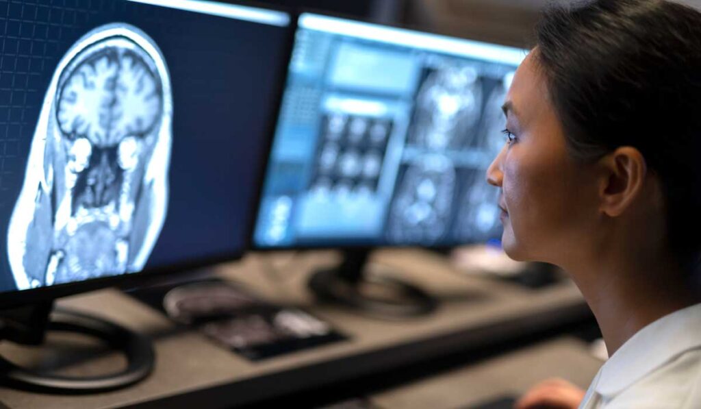
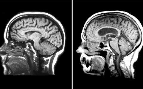
Offering a high-resolution view into the human brain, powerful 7-Tesla (7-T) magnetic resonance imaging (MRI) is on track to improve the diagnosis and treatment of neurological disorders by leaps and bounds.
While exciting, this development comes as no surprise to neuroimaging expert Francesca Bagnato, M.D., associate vice chair for research at Vanderbilt University Medical Center.
Almost 10 years ago, Bagnato published one of the first studies to demonstrate the utility of using 7-Tesla MRI to detect microglia activation, a pathological change associated with multiple sclerosis (MS). The high-definition technology was FDA-approved in 2017 for neurological imaging and other medical uses.
With funding from the National MS Society, Bagnato and colleagues in the MS Center at VUMC and the Vanderbilt University Institute of Imaging are investigating whether 7-T MRI, as well as other state-of-the-art MRI techniques, can be used to identify and monitor early signs of MS neurodegenerative disease and predict the likelihood of severe progression.
A Complex Interplay
Commonly, clinicians consider relapsing-remitting MS to be the initial form that eventually degenerates into secondary progressive MS, Bagnato said. During the relapsing-remitting phase, acute inflammation predominates, whereas secondary progressive MS is sustained by neurodegeneration and microglia activation.
In contrast, Bagnato thinks all three processes affect patients at any stage of the disease. “It’s just a matter of which one predominates when,” she said, “but in the end, the result derives from their interplay.”
“The relationships between these three main pathological processes – inflammation, neurodegeneration and microglia activation – are very complex, and likely not homogeneous across the board,” Bagnato explained. “At this time, we have no ability to predict early in the disease which process will be the main culprit of the disease.”
Paramagnetic Rims
Bagnato’s initial research with 7-T imaging started due to an interest in iron.
“The relationships between these three main pathological processes – inflammation, neurodegeneration and microglia activation – are very complex, and likely not homogeneous.”
For unknown reasons, patients with MS display alterations in iron abundance in the brain, including a specific increase in iron stored by microglia. Employing the sensitivity of 7-T MRI, Bagnato and colleagues discovered that some MS lesions are surrounded by a notable ring of iron (later termed paramagnetic rims), indicative of activated microglia.
“These lesions harbor a core of profound demyelination,” Bagnato explained. “They are chronic active, slowly expanding over time. Then at some point, as the microglia become dormant, they stop expanding and transform into chronic inactive lesions featured by profound tissue loss.”
MRI Biomarkers
As part of a new research project, Bagnato is using 7-T MRI to identify paramagnetic rims as they develop in patients newly diagnosed with MS. She aims to assess whether detection of these iron rings early in the disease course is predictive of worse outcomes.
A major strength of the approach, Bagnato explains, is that paramagnetic rims would make for an easily detectable biomarker.
“We don’t want our radiologists to have to spend hours just to read one MRI of an MS patient,” she said. “Lesions with paramagnetic rims have distinctive features, allowing a radiologist to say, right away, if they are present or not.”
Translating the Technology
Due to their novelty and expense, 7-T MRI systems are only available at VUMC and a limited number of other medical centers, and the majority are used strictly for research. Bagnato does not see this as a limitation but as an opportunity to translate the 7-T MRI methodology to the more widely available 3-T MRI systems.
“What the 7-T has really taught us is that by refining MRI sequences, we can see more,” Bagnato said. “It’s very possible that by using the 7-T derived knowledge to manipulate the sequences that we have available at 3-T, we are going to be able to discover more about disease in general.”


