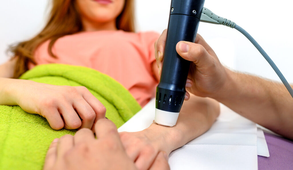
While standard radiography has long been the go-to diagnostic tool for rheumatologic diseases, the advent of musculoskeletal (MSK) ultrasonography opens the door to earlier detection.
This shift has had a significant impact on inflammatory forms of arthritis that can cause permanent physical damage over time.
“One of the main drawbacks of conventional imaging is the inability to detect early changes in the connective tissue that lines the joints and other periarticular tissues,” said Erin Yee-May Chew, M.D., a rheumatologist at Vanderbilt University Medical Center.
An MSK ultrasound offers quicker and more accurate diagnoses, a chance to optimize treatment, and more accurate needle placement for therapeutic joint injections and aspirations, Chew says. Its use has proven beneficial in several rheumatologic diseases, including gout, rheumatoid arthritis (RA), psoriatic arthritis, as well as conditions such as lupus and scleroderma.
Beyond Standard Imaging
Magnetic resonance imaging (MRI) is often preferred for detection of soft tissue pathologies and early bony inflammatory changes, yet its application has been limited.
“The financial burden of MRI deters its general use,” Chew said.
Similar to an MRI, ultrasonography is noninvasive and does not involve exposure to ionizing radiation. Yet, it is more economical and portable than MRI, with the ability to image tendons under different levels of stress through dynamic examinations.
“Advancement of transducer technologies has made it possible to image smaller, superficial MSK structures with excellent resolution,” Chew said.
The technologies have been transformative for MSK imaging, especially when examining small and difficult-to-reach body parts. Chew notes standard radiography often fails to detect early changes in soft tissue, like synovial proliferation in RA or shifts in cartilage architecture in early osteoarthritis.
Applying Expertise
In the case of RA, MSK ultrasound can be used for early disease detection, to guide treatment decisions, monitor disease progression, and for follow-up.
“This modality allows us to make definitive diagnoses very quickly,” Chew said. “We can perform and interpret an ultrasound as an extension of our history and physical exam on the same day of the patient visit, determining if there is active inflammation in the joints that requires immediate treatment.”
“This modality allows us to make definitive diagnoses very quickly.”
Owing to the complex nature of MSK ultrasound, Chew says it is imperative for trained specialists to assess pathologies. Knowledge about the technical aspects of the machine, as well as familiarity with the transducer and the artifacts is essential.
Chew was the first rheumatologist at Vanderbilt to complete advanced ultrasound training in the Ultrasound School of North American Rheumatologists program and to obtain the MSK Ultrasound Certification in Rheumatology, the professional certification for clinicians who perform ultrasound as part of their practice in rheumatology.
She said she intends to use her expertise to develop a specialized ultrasound program in the Division of Rheumatology and Immunology at Vanderbilt.
Roadmap for the Future
Chew says the increasing significance of MSK ultrasound in the diagnosis and management of several rheumatological conditions may impact physician training and education, payments and reimbursement, as well as competencies and certifications.
As the technology continues to evolve, she expects its applications will extend beyond existing diagnostic and interventional approaches, enabling the creation of new uses.
“Alongside Tracy Frech, M.D., I’m designing a new protocol for evaluating hand pain using MSK ultrasound in patients with systemic sclerosis,” Chew said. “I’m excited about the future of this technology and the possibility of having a small impact on its direction.”


