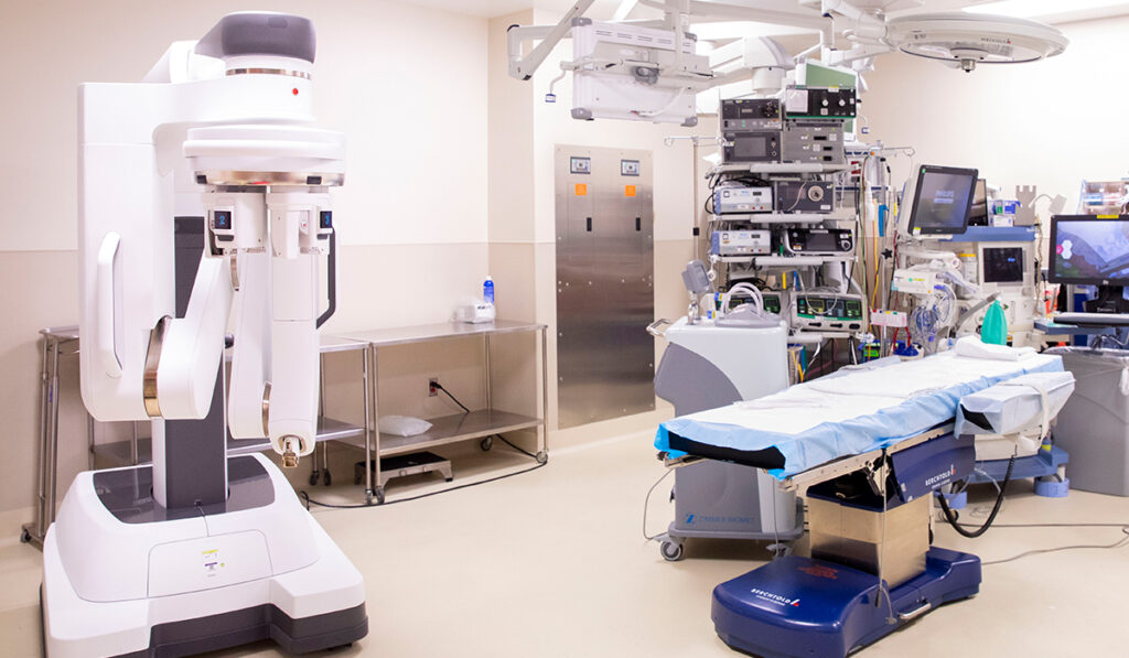
In what may be the first of its kind, surgeons at Monroe Carell Jr. Children’s Hospital at Vanderbilt used a robotic-assisted minimally invasive technique to repair a congenital rectourethral fistula in a 2-year-old boy with a urethral duplication.
This form of anorectal malformation occurs rarely, so the repair involved decisions made in somewhat uncharted territory. Typically, they said, a surgery of this complexity in a small child would be open, but superior deep pelvic visualization as well as advanced instrumentation were the deciding factors on attempting the repair robotically.
With Vanderbilt and Monroe Carell at the forefront of pediatric robotic surgery, John Thomas, M.D., a professor in the Division of Pediatric Urology, and urology chief resident Rohan Bhalla, M.D., tackled this laparoscopic transperitoneal rectourethral fistula repair on the smallest patient they have operated on robotically to date.
“This is such a rare anomaly, but the robotic approach for accessing deep in the pelvis is well-known,” Thomas said. “This approach gave us superior visualization of all-important structures and allowed for resection of this child’s very deep fistulous tract without complications.”
Open vs. Robotic
The Monroe Carell team has primarily used robotics in reconstructive kidney, ureteral, and bladder surgeries, sometimes in children as young as six-months old.
“When you compare robotics in kids versus robotics in adults, you have to realize that kids seem infinitely smaller,” Bhalla said. “That means that your ability to put ports in and insufflate the patient so that you can see pertinent anatomy is challenging. The open approach sometimes tends to be better because you can get better exposure to the abdomen and pelvis.”
Most young pediatric patients do well no matter the approach, Bhalla said.
“We chose to do it robotically, knowing that if we ran into significant adhesions or other barriers, we could convert to open surgery.”
The dexterity made possible through the da Vinci XI was one of the deciding factors, Thomas added, “since visualizing tissues deep into the pelvis to resect and suture was necessary.”
Case Synopsis
At age three months, the boy had a routine voiding cystourethrogram to evaluate a febrile urinary tract infection and watery stools, but the rectourethral fistula was not apparent through this test. Close collaboration with pediatric general surgery helped guide further testing.
A CT cystogram later identified the fistula tract and enabled diagnosis of bilateral bladder diverticula and urethral duplication with rectourethral fistula. The patient had a dominant ventral meatus with a pinpoint accessory dorsal meatus, and an orthotopic anus. The accessory urethra communicated proximally at the verumontanum.
The young patient had a full connection between the rectum and the dominant urethra as well as the duplicate urethra. This left him vulnerable to infection, incontinence, or stool in the urinary tract, potentially infecting the kidneys.
The repair began with a hitch stitch through the bladder to expose the peritoneal reflection. Once the fistula was located, the tract between the rectum and posterior urethra was divided and the duplicated urethra was ligated. The rectal defect was then closed and the peritoneum advanced to cover the site of the fistula.
Safe and Efficacious
The outcomes were as hoped. During the 229-minute procedure, blood loss was minimal. The child was discharged on post-operative day three after a negative barium enema. Two weeks later, his catheter was removed, and he was voiding normally and having regular bowel movements.
“I may never see this specific problem again in my career, so the fact that this child did well is a testament to all involved.”
“I may never see this specific problem again in my career, so the fact that this child did well is a testament to all involved, who were willing to apply a different technique that turned out to be the right one for this patient,” Thomas said. “We were able to conclude that this is a safe and feasible approach with minimal morbidity that allows for excellent visualization, avoids an open laparotomy and potentially a staged procedure.”
Expanding Adoption
Monroe Carell is a high-volume pediatric center that attracts complex cases like this one. Collaborating with the adult robotic surgeons has resulted in a rapid expansion of the technology, breaking through new barriers, year by year. Along with urology, pediatric general surgery under the direction and efforts of Irving Zamora, M.D., has adopted robotic techniques in their patients over the last two years.
“These kinds of robotic surgeries of the rectum, bladder and urethra are something that we do on the adult side very frequently,” Bhalla said. “But in pediatrics, we are still near the starting block. Cases like this are encouraging for extending this to more of our younger patients.”


