As recently as a decade ago, ultrasound technology could not effectively guide surgeons through minimally invasive laparoscopic procedures for adults, much less pediatric patients.
Now, following the adult hospital’s lead, Monroe Carell Jr. Children’s Hospital at Vanderbilt has acquired both the technology and the skills to conduct ultrasound-guided minimally invasive procedures, and a recent case study reports on one outcome.
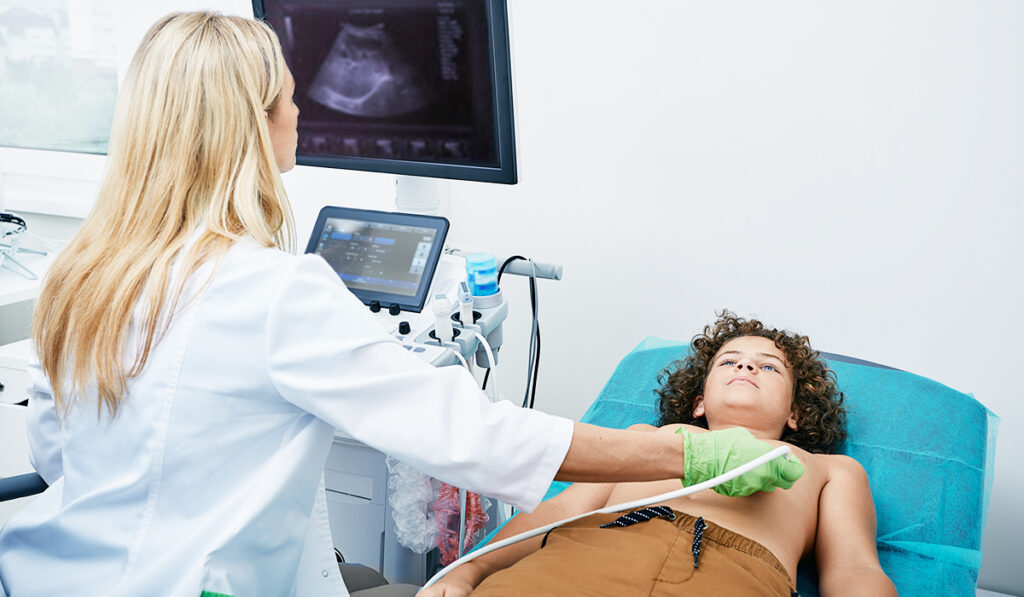
“We have been using intraoperative ultrasound for some time now, but only in open operations,” said pediatric surgeon Irving Zamora, M.D. “This new technology produces really clear, crisp images, using a 10 mm probe, which allows us to use it laparoscopically.”
A Test Case
While Zamora and his colleagues have discovered myriad applications for the technology, they recently put it to the test in a complex spleen-preserving distal pancreatectomy case involving a 17-year old boy with multiple endocrine neoplasia (MEN).
“In this patient, we were able to use laparoscopy to safely visualize the pancreas, one of the most challenging organs to access,” Zamora, who worked with Vanderbilt pediatric surgeon Harold N. Lovvorn, M.D., on the case.
“Intraoperative ultrasound enabled us to identify all the lesions and their relationship to the vessels in order to perform a very precise surgical resection in a minimally invasive fashion. This led to an amazing outcome, because he recovered very quickly. We were able to preserve the remainder of the pancreas and other important surrounding structures, mainly the vessels. We were also able to preserve the spleen, which is really important in a child who could potentially become immunocompromised.”
Challenge Met
The patient had been followed for some time by Monroe Carell endocrinologist, Karishma Datye, M.D., who tracked lesions in the parathyroid gland, pituitary gland and pancreas, the primary structures MEN affects.
Datye reached out to Zamora, describing the pancreatic lesions and a strong family history of MEN. Together, they discussed whether or not to remove the lesions.
“We monitored the patient with serial MRI and laboratory work for about a year trying to ascertain whether these lesions were active, but what we couldn’t know is if they were destined to become malignant or not,” Zamora said. “We finally decided the risk of malignancy was high enough that it was in his best interest to resect them.”
“We knew we had clear surgical margins and that we were sparing vasculature and other structures.”
Through a 12 mm incision, the team utilized a new BK intraoperative ultrasound probe to identify the five different lesions inside the pancreas and applied color Doppler to identify the vessels and move the probe across the surface of the pancreas, enabling real-time identification of the lesions.
“MRI tells us where the lesions are, but when you perform the actual surgery, those images alone do not give you optimal confidence that you’re identifying and removing all of them,” Zamora said. “This way, we knew we had clear surgical margins and that we were sparing vasculature and other structures,” he said.
Watchful and Ready
The young man’s lesions were not malignant. He recovered quickly and returned home two days post-surgery.
The endocrinology team is currently monitoring a lesion on his pituitary gland and monitoring his parathyroid glands and remaining pancreas through serial imaging and laboratory work to see if he needs further interventions.
“I believe the fact that he knew these lesions were not malignant, along with knowing that we are remaining vigilant to identify changes in the other vulnerable organs early has allowed him to breathe easier,” Zamora said. “This is another huge benefit of our more precise, patient-specific management approach to this case.”
The team is also following the patient’s brother, who has some lesions and may end up with the same procedure.
Other Applications
Zamora attributes the case’s success largely to the new BK Medical ultrasound equipment, obtained with support from donors to Monroe Carell’s growing minimally invasive cancer surgery program.
But he also emphasized a growing skillset among surgeons in the department to conduct laparoscopic surgery in children.
“Our capabilities with advanced minimally invasive surgery in children continues to grow as we gain expertise and experience,” Zamora said. “This was the first time this kind of procedure had been performed on a pediatric patient here at Vanderbilt. We will continue increasingly to try to employ precision image-guided surgery for our patients.”