A team of investigators at Vanderbilt University Medical Center has recently developed and validated a radiomics-based machine-learning model to classify musculoskeletal myxoid soft tissue tumors.
“Distinguishing benign and malignant myxoid tumors can be challenging due to their overlapping clinical, imaging, and histologic features. Based on our clinical data, the rate of indeterminate preoperative diagnosis of myxoid tumors is around 30 to 40 percent,” explained Joshua M. Lawrenz, M.D., an assistant professor in the Division of Musculoskeletal Oncology at Vanderbilt.
“Because treatment strategies differ between the two diagnoses, an accurate preoperative diagnosis is critical.”
Radiomics-Based Model Development
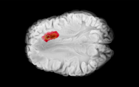
To assess whether radiomics could help improve the ability to diagnose tumors by providing more information from MRI studies, Lawrenz and colleagues established a multidisciplinary collaboration between orthopaedic oncology, musculoskeletal radiology, biostatistics, and the Vanderbilt Institute for Surgery and Engineering (VISE).
The investigators conducted a retrospective review of 90 patients with a pretreatment MRI and a histologically confirmed myxoid soft tissue tumor, of which 45 were benign and 45 were malignant. Baseline clinical features for patients — including patient age, sex, tumor size, tumor depth, tumor location and pain — were used in the review.
Attending clinicians performed manual image segmentation of tumors, which allowed for the extraction of over 100 radiomics features, pertaining to their shape, intensity and texture. These features, along with clinical features, were used to train five different machine-learning models to classify tumors as benign or malignant in an initial cohort of 40 patients. The team then validated the models’ predictive ability using a separate cohort of 50 patients.
Head-to-Head with Radiologists
The Vanderbilt team selected the best classifier model using the validation cohort and compared its findings with those from images reviewed by musculoskeletal radiologists. Two attending radiologists were provided the same images and baseline clinical features for each tumor. They independently classified the 50 tumors in the validation cohort as benign or malignant. When their diagnoses were discordant, a third senior radiologist made the consensus diagnosis.
The findings, recently presented at the annual meetings of the American College of Surgeons and the Musculoskeletal Tumor Society, demonstrated that the machine-learning model accurately predicted 78 percent of tumors correctly – a rate similar to that of the consensus imaging interpretation by three musculoskeletal radiologists, which came in at 74 percent.
Importantly, the researchers found that the radiologists’ performance correlated with their confidence in the diagnosis. When radiology rated their confidence as high, they were 95 percent accurate. When they rated their confidence as intermediate or low, they were 62 percent accurate. These data suggest that radiomics may provide diagnostic aid when radiologist confidence is low or when radiologists disagree on the diagnosis.
“I can’t overstate how outstanding our musculoskeletal radiologists are. And what’s neat about their collaboration in the design and execution of this study is their desire to learn how we can use this advanced technology to augment our understanding of images even more, potentially allowing us to make more accurate diagnoses and better treatment decisions,” Lawrenz said.
Future Directions
The Vanderbilt team aims to validate the model in a larger, external cohort with the hope to develop an easy-to-use tool that could be tested prospectively in the clinical setting.
The investigators also envision this research could be applied to other clinical scenarios in musculoskeletal oncology, such as in the diagnosis of indeterminant pulmonary nodules or local recurrence.


