Ultrasound for inflammatory bowel disease (IBD) has taken a back seat since the increasing and widespread availability of MRI and CT over the past few decades.
Now, gastroenterologists are recognizing that the current generation of ultrasound machines with high-resolution imaging provides valuable information on the state of current disease activity, and the modality is beginning to make a comeback.
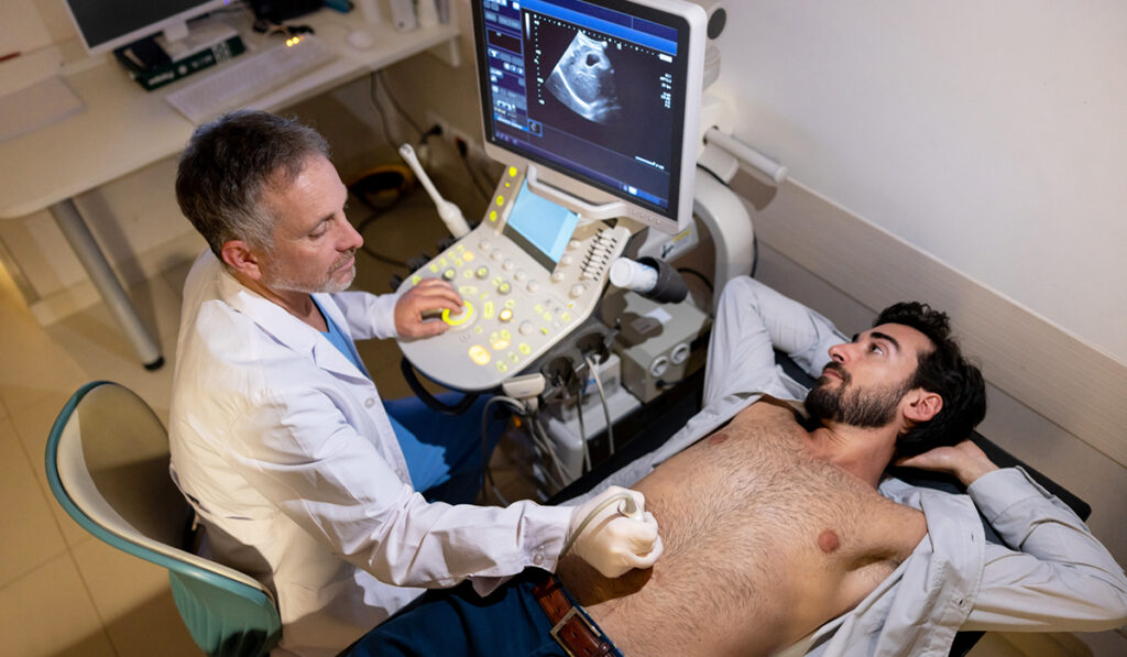
Gastroenterologist Baldeep Pabla, M.D., at Vanderbilt University Medical Center, was early to embrace these new capabilities and become certified in point-of-care abdominal ultrasound in 2021. David Schwartz, M.D., director of the IBD Clinic at Vanderbilt, was certified in October 2023. Together, they are among the pioneers providing patients with same-day diagnoses and treatment plans.
“Since patients with IBD have ebbs and flows in their inflammation, a patient-friendly, accurate means of detecting and monitoring this is the name of the game.”
“We acquire the images, interpret them, and make treatment decisions all within one visit; that’s a really unique way to measure and monitor inflammation,” Pabla said.
Since 2021, Pabla has performed over 450 point-of-care ultrasounds and has presented an American Gastroenterology Association abstract demonstrating its high sensitivity and specificity in moderate and severe ulcerative colitis and Crohn’s disease. He is now consolidating patient-satisfaction survey data for publication.
“Since patients with IBD have ebbs and flows in their inflammation, a patient-friendly, accurate means of detecting and monitoring this is the name of the game, and ultrasound is a viable way to accomplish that,” he said.
Point-of-Care Benefits
To evaluate inflammatory activity at the time of the patient visit, Pabla measures the thickness of the bowel wall throughout the colon, and of the small bowel, with a focus at the terminal ileum, a portion of the bowel that is commonly involved in Crohn’s disease. He also runs a color Doppler signal to look for visible blood flow in the mesentery tributaries, applying the modified Limberg grading scale to determine, on a scale from 0 to 3, how active the disease appears in that segment.
“In a healthy bowel with non-contrast enhanced ultrasound, you really shouldn’t see the Doppler signal in these tributaries, so when you do, that can speak to significant inflammation and disease activity,” Pabla explained.
He adds that blood and stool tests have good sensitivity and specificity for inflammation but cannot supply the information on different grades and locations of inflammation that imaging does. A point-of-care ultrasound is a less expensive and more patient-friendly alternative to endoscopy and these serum and stool studies. It also avoids ionizing radiation used in CT and MRI.
Pabla does not discount the need for endoscopy in IBD, particularly for tissue sampling in patients with longstanding colitis to test for dysplasia and precancerous lesions.
“Endoscopy with biopsies is ultimately needed for these patients, but ultrasounds may defer and lower the frequency of these invasive procedures. In other patients, though, like those with mild Crohn’s disease or negative results for inflammatory disease in past testing, ultrasound exams may drastically reduce the need for endoscopy,” he said.
Future Studies
One of the applications Pabla is also looking to examine is in the management of acute, severe ulcerative colitis in patients admitted to the hospital.
“There are ongoing studies looking at scanning those patients serially almost every day to determine whether they’re responding to therapy or not,” he said.
This information can help doctors decide whether to proceed to surgery, such as colectomy, or switch to other therapies.
A second area of exploration is ultrasound’s ability to evaluate transmural healing in Crohn’s disease patients, looking at whether the bowel wall has returned to normal after treatment, Pabla said. These studies also will assess whether ultrasound markers may be better than inflammatory markers for indications that patients are doing well over the long term.
As yet another focus, shear wave elastography, a high-intensity ultrasound application that is gaining traction in lesion and nodule testing, might discern between an inflammatory and a fibrotic stricture in the intestinal tract. This would guide treatment decisions on whether surgery, which is indicated only for fibrotic strictures, is warranted.