At 213 per 10,000 births, the rate of congenital heart disease in the state of Tennessee is one of the highest in the country, and Monroe Carell Jr. Children’s Hospital at Vanderbilt sees some of the most complex cases from across the Southeast at its fetal surgery center.
It may not seem remarkable, then, that pediatric cardiology specialist David A. Parra, M.D., director of advanced cardiac imaging, receives frequent referrals for infants with rare cardiac conditions. One of these was a male newborn with a sizable cardiac mass discovered on prenatal echocardiography.
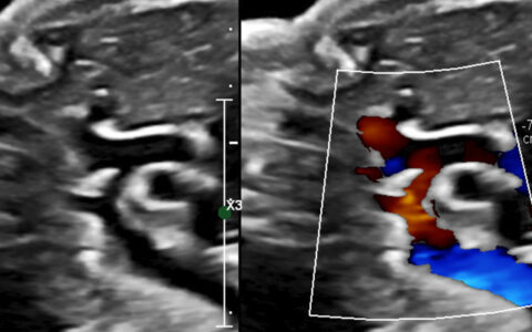
Information gathered initially led toward a diagnosis of rhabdomyoma, but careful inspection by color Doppler revealed blood flow that confounded that conclusion, Parra said. Other potential causes were eliminated and the diagnosis became a rare pseudoaneurysm of the mitral aortic intervalvular fibrosa. This triggered treatment with aspirin and a follow-up regime to monitor the mass’s growth or shrinkage.
“This case underscores the importance of keeping this diagnosis in consideration for atrial masses noted on fetal echocardiography,” he said. “Doing so puts clinicians in a better position to directly counsel families prenatally and guide postnatal management.”
Multiple Possibilities
It was also better news for the parents. The referring obstetrician, not yet detecting blood flow at 31-weeks’ gestation, had concerns the patient was forming rhabdomyoma tumors, which are often associated with tuberous sclerosis. Patients with this condition can develop multiple benign tumors at various sites in the body. Only 30 percent of rhabdomyomas present as single tumors, so the finding would carry an expectation of additional masses developing over time.
A pseudoaneurysm occurs when a weakened blood vessel wall in the myocardium fails as a result of a direct injury or congenitally, as in this case. Unlike a rhabdomyoma or other tumor, a pseudoaneurysm has blood flow within it. When that blood flow is undetectable, it may mimic a pseudoaneurysm or a true aneurysm, which involves a breach in the innermost layer of an artery.
This Case
The mother of the newborn had an unremarkable pregnancy, despite a history of supraventricular tachycardia managed on metoprolol. At the 31-week echocardiogram, the infant had an 8- to 10-mm echogenic mass in the atrium, with a neck that extended from the aortic valve ring. The fetal rhythm was normal, and there was no obstruction of the ventricular inflow or outflow tracts.
A mass found in the flap of the septum primum was consistent with a rhabdomyoma, but the specific site – in the region of mitral-aortic continuity – was not. It was the blood flow Parra detected by post-natal color Doppler examination that made a differential diagnosis possible.
“Both the location of the mass and its connection to the left ventricular outflow and to-and-fro blood movement can be used to distinguish this diagnosis from tumors or other potential etiologies,” Parra said. “Further, this mass expanded with systole, collapsed with diastole. This limited the possibilities to aneurysm or pseudoaneurysm, but its location and shape were not consistent with a true aneurysm.”
The infant has thrived and is asymptomatic at 3 years old. He receives daily aspirin to avoid formation of clots in the mass that might migrate to the aortic outflow tract.
“He continues to be monitored conservatively with echocardiography and clinical assessment, which we think is appropriate in the absence of other findings or clinical symptoms,” Parra said.
Optimized Visibility
Parra says postnatal imaging was done with higher frequency imaging than is possible with fetal echocardiography, enabling superior visualization of both 2D images and 3D color.
“When needed, other imaging modalities, such as transesophageal echocardiography, cardiovascular computed tomography and magnetic resonance could be used postnatally to further diagnose and characterize the mass, including its relationship to other cardiac structures,” Parra said.
“In this case, our transthoracic echocardiography did the job.”


