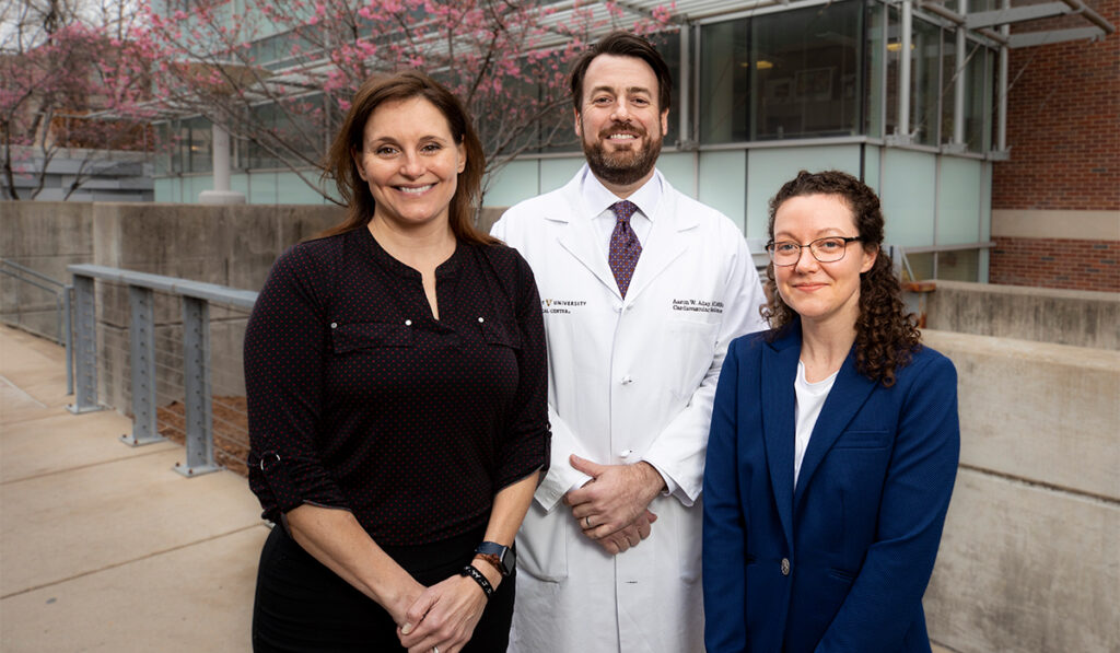
Rachelle Crescenzi, Ph.D., Aaron Aday, M.D., and colleagues at Vanderbilt University Medical Center are transforming the diagnosis and treatment of lipedema.
The painful condition, often confused with obesity, is caused by an abnormal deposition of fatty tissue and almost exclusively affects women – an estimated 11 percent worldwide. Despite its prevalence, diagnosis remains a challenge, and treatments are limited.
“It wasn’t thought that you could compress out any of the pathologic tissue in lipedema, that it was just fat.”
To address this, Crescenzi and colleagues have developed MRI strategies to quantify tissue sodium and fat content throughout the body, finding that the measurements can distinguish patients with lipedema from those with a similar BMI but without the condition.
Physical therapy may then be applied to correct tissue sodium levels and improve lipedema-related pain and mobility, the team reports.
“It wasn’t thought that you could compress out any of the pathologic tissue in lipedema, that it was just fat. But it really is fat and edema,” Crescenzi said. “We think sodium is a marker of inflammation, and it reduces after therapy.”
To further advance this work, Crescenzi and Aday were recently awarded over $700,000 from the Lipedema Foundation to establish a biorepository focused on lipedema. Previously, in 2021, Crescenzi received the first R01 ever awarded for lipedema research by the NIH.
Distinguishing From Obesity
While often mistaken for obesity, lipedema fat is resistant to diet or exercise. Patients experience an accumulation of fluid in addition to fat, putting them at risk for complications.
“Excess fluid could contribute to inflammation in affected areas, for instance,” Crescenzi said.
In women, the accumulation of fat occurs mainly in the legs, causing them “a lot of pain and difficulty with their daily activities,” said Aday, a vascular medicine specialist at Vanderbilt.
“It’s a real disorder,” Aday said. Yet many women “are having a horrible time, trying to find a diagnosis.”
About seven years ago, Crescenzi, then a postdoctoral fellow in the Department of Radiology and Radiological Sciences at Vanderbilt, began to apply imaging techniques to improve diagnosis.
Lipedema was initially characterized in the 1940s, but the diagnosis relied on external measurements, which made it challenging, Crescenzi said.
With imaging, investigators can peer inside the body, seeing differences between lipedema and obesity, she added.
Benefits of Conservative Therapies
Conservative therapy tactics included exercise guidance, nutrition, education, compression and manual therapy. The manual techniques use lymphatic drainage massage to impact deep and superficial tissues, myofascial and soft tissue releases to relax contracted muscles and surrounding connective tissues, and graded negative pressure to expand and stretch tissue — all of which can mitigate pain and optimize lymphatic circulation.
“Manual therapy paired with a tailored exercise and self-management program is very effective in changing their quality of life.”
“The patients received a full physical therapy, patient-tailored approach,” said Paula Donahue, D.P.T., a Lymphology Association of North America-certified Lymphedema Therapist at Vanderbilt.
Donahue has shown that in women with early lipedema, physical therapy techniques act directly on the source of their pain, resulting in improved function and reductions in tissue sodium in the skin and fat as measured by MRI.
A patient dealing with unexplained pain that her doctor didn’t know how to address found total relief with physical therapy, Donahue said.
“For these individuals, manual therapy paired with a tailored exercise and self-management program is very effective in changing their quality of life.”
Though not included in their published pilot study, the importance of interdisciplinary care to address additional health needs, such as nutritional counseling, medical or surgical weight-loss, lipedema-reducing liposuction and psychological support is something Donahue stresses.
Developing a National Resource
While Crescenzi’s initial focus has been on MRI technologies, she and colleagues are also testing whether lower-cost ultrasound may also be used to quantify the extent of abnormal fat accumulation in the legs of patients with lipedema.
“We’re in the infancy of understanding this disease.”
The team also is applying magnetic resonance lymphangiography to better understand the hallmark features of lipedema.
“We think the vascular system is overloaded with edema, and it shows up on angiography,” Crescenzi said.
“We’re in the infancy of understanding this disease,” said Aday, co-author of the team’s paper on the angiography technique published in the Journal of Magnetic Resonance Imaging.
By hosting well-characterized biological samples from individuals with and without lipedema, the newly-funded biorepository will fuel research into the underlying causes of the condition, leading to advances in treatment and prevention.
“We’re in a position to develop a national resource for lipedema,” Crescenzi said.


