Fastidious follow-up with patients who have undergone hematopoietic stem cell transplant (HSCT) is critical to responding with timely interventions to increase survival and lower morbidity.
In a serendipitous discovery, researchers found that the movement of leukocytes along the walls of a patient’s microvessels tells an important story about their likelihood for relapse and overall survival after HSCT.
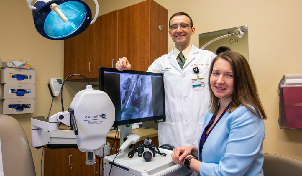
A Vanderbilt University Medical Center team, led by dermatologist Eric Tkaczyk, M.D., founding director of the Vanderbilt Dermatology Translational Research Clinic, and adjoint assistant professor Inga Saknite, Ph.D., tested reflectance confocal microscopy (RCM), a technology used for years as a substitute biopsy for certain kinds of suspicious skin lesions. Their goal was to see if it would predict outcomes in patients after HSCT.
Their findings, published in JAMA Dermatology, demonstrated a high association between patient outcomes and the leukocyte-endothelial interactions visualized directly in tiny, upper dermal capillaries after HSCT.
“This 10-minute video scan of the skin was a much stronger predictor for patient outcomes than the best existing clinical-risk assessment,” Saknite said.
“Up to 70 percent of all deaths after HSCT are associated with relapse,” Tkaczyk said. “Assessing and monitoring this dynamic marker after transplant could help guide interventions for patients at high risk for relapse.”
A Keen Eye
RCM has been used for years as an “optical biopsy” for certain kinds of suspicious skin lesions. In RCM, the light reflected from a specific depth, typically about 0.1 millimeters below the skin surface, is collected to create a high-resolution, magnified image, Saknite explained.
“This 10-minute video scan of the skin was a much stronger predictor for patient outcomes than the best existing clinical-risk assessment.”
Using RCM, Tkaczyk observed the well-defined movement of leukocytes inside capillaries, noting a laminar flow in some patients and more turbulent flows in others. Sometimes, the movements would occur in an erratic manner before sticking to the endothelial wall.
Curious to see if RCM inspection would reveal whether patients with graft-versus-host-disease (GVHD) after HSCT had different, more inflammatory biomarkers than those without GVHD, Tkaczyk and Saknite conducted a cross-sectional study to examine differences in leukocyte behavior. They imaged the skin of 10 patients in each group but found no better sensitivity for GVHD than from conventional biopsy.
However, Saknite began to observe an association between poor survival and a high rate of rolling and sticking leukocytes.
“I thought it was peculiar: Why do some of these post-transplant patients, even without GVHD, have this particularly inflammatory response of increased immune cell activity?” Saknite asked.
Resounding Results
To find an answer, the team with two medical students conducted a cohort investigation, examining thousands of RCM videos on 56 patients who underwent HSCT. After following the patients for 100 days, the researchers discovered that three times as many patients with the abnormal leukocyte behavior relapsed and died compared to those with normal flow.
“While we can’t fully explain why, the extent to which the leukocytes roll and adhere is predictive of worse outcomes,” Saknite said.
This was an auspicious finding, as the discovery of biomarkers to predict relapse after HSCT has been elusive.
In the HSCT Clinic
After HSCT, patients are routinely monitored with bone-marrow biopsies, usually around day 30 and 100 post-transplant. Using RCM, however, may more easily shed light on biomarkers of poor response. In addition, Saknite said the procedure could help identify patients who might take a turn for the worse before there is clinical evidence of relapse based on bone marrow biopsy.
Tkaczyk believes this approach may help physicians directly quantify intact immunological processes at the patient’s bedside, since clinicians “walk a tightrope when it comes to how much immunosuppression to induce through drugs.”
Knowing that a patient is having an inflammatory response at the microcirculatory level allows proactive intervention. It may be that disease relapse is imminent or that the immunosuppression dosing needs to be lowered. This type of knowledge could give HSCT clinicians the chance to investigate and intervene, potentially even with an early second transplant or salvage therapy.
In addition, patients at high risk for relapse could be more readily referred to clinical trials.
Validation and Future Development
The team is looking toward a larger, multicenter clinical trial to validate their findings. Saknite is also hoping vascular biologists will take on the challenge of furthering the investigation.
“First, we need a vascular biologist to dive into the science of this and explain the mechanism of this response in HSCT more completely,” Saknite said. “Then, I think we can move forward with a multicenter study.”
This work may ultimately lead to applications for a variety of inflammatory diseases, including autoimmune conditions, where immunosuppression levels also must be weighed against vulnerability to infection.


