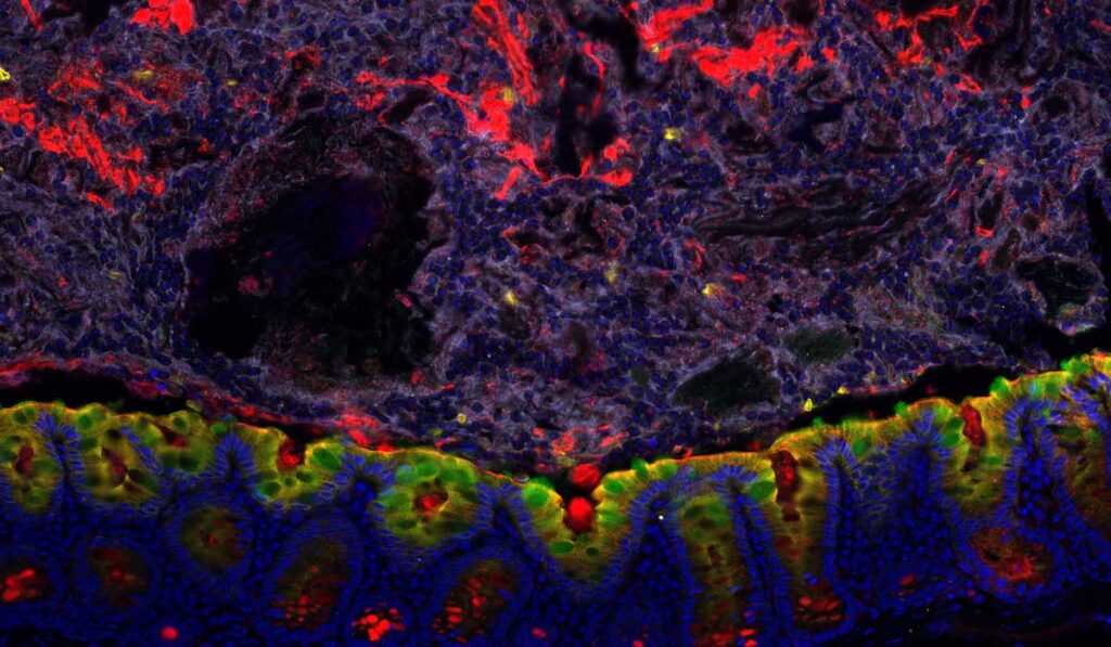
A new clinically adaptable polymer developed by investigators at Vanderbilt University Medical Center enables dual visualization of colonic tissue and mucosal-associated microbes. While techniques exist to preserve and examine either tissue or microbes separately, the new approach addresses a pressing need for histopathology workflows that permit simultaneous analysis of host cells and accompanying bacteria.
“This fixation method enables the study of complex interactions occurring between cells and gut microbes in clinical specimens,” said Ken Lau, Ph.D., lead author on the study and an associate professor of cell and developmental biology at Vanderbilt. “We piloted the method at Vanderbilt to ensure that there’s no barrier to incorporation into clinical pipelines.”
“The idea is we just take the Poloxamer solution out of the refrigerator, fix the sample in that solution, and let it get to room temperature, that’s very attractive and applicable to clinical pipelines.”
The original method, recently published in npj Biofilms and Microbiomes, is currently being applied to the large-scale effort at Vanderbilt to build a spatial atlas of precancerous lesions in the colon, part of the National Cancer Institute’s Human Tumor Atlas Network.
“We want to have a multiplex characterization of both the microbes and the host cells in the colon so that we can look at potential interactions between the two during the process of carcinogenesis,” Lau said.
Mucosal-associated Biofilms
In the healthy colon, two layers of mucus blanket the intestinal epithelium. While the outer layer is routinely colonized by microbes, the inner mucus layer, located directly above the epithelial surface, is relatively sterile. When the sterility of the inner mucus layer is breached, mucosal-associated biofilms may form, and these biofilms are associated with the development of colorectal cancer.
“We want to have a multiplex characterization of both the microbes and the host cells in the colon so that we can look at potential interactions between the two during the process of carcinogenesis.”
“Mucosal-associated biofilms are colonies of microbes that form a self-organizing community on the surface of the mucosa,” Lau said. “These microbes are in direct contact with the intestinal epithelium and can create their own microenvironment, which will affect the function of surrounding cells.”
Poloxamer 407 Samples Superior
While there is intense interest in the role of the gut microbiome in colorectal cancer and gastrointestinal disease, the critically important colonic mucus in which pathogenic microbes reside is readily dissolved by the standard formalin-fixed, paraffin-embedded (FFPE) tissue preservation method.
“The FFPE method enables visualization of tissue architecture and morphology, but you lose all information with regards to the spatial organization of the mucus and the microbes associated with the mucus,” Lau explained. Another commonly used fixative, Carnoy’s solution, preserves colonic mucus but reduces the sensitivity of immunofluorescence and fluorescence in situ hybridization (FISH) necessary for the detection of host factors.
“Our goal was to devise a strategy so that we can assess the spatial organization of the mucus and the microbes together with the host cells in a multiplex fashion,” Lau said.
In the new study, Lau and his team demonstrate that Poloxamer 407-fixed samples are superior to Carnoy’s-fixed samples for visualizing both mucosa-associated microbes and host cells in the same tissue section. Furthermore, Poloxamer 407 polymerizes at room temperature, enabling its easy incorporation into clinical protocols.
“The idea is we just take the Poloxamer solution out of the refrigerator, fix the sample in that solution, and let it get to room temperature,” Lau explained. “That’s very attractive and applicable to clinical pipelines.”
Discoveries Ahead with Multiplex Imaging
The novel fixation method enabled Lau and colleagues to integrate multimodal analysis using a modified multiplex immunofluorescence protocol.
“Our efforts represent an advance in the field of multiplex imaging, and one of the first to simultaneously evaluate composition and organization of both microbes and host cells,” wrote the authors. “Methods that enable multimodal analysis will become very valuable in future endeavors for understanding complex tissue ecosystems.”


