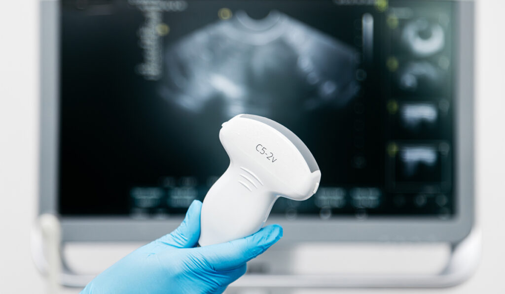
While computed tomography and magnetic resonance imaging have long been the go-to postoperative imaging tools for patients with Chiari malformations, the advent of transcutaneous ultrasonography opens the door to inexpensive, radiation-free, point-of-care assessment.
“One of the main drawbacks of conventional static imaging is the inability to assess changes in different head positions and body postures,” said Ryan P. Lee, MD, a neurosurgeon and director of the Hydrocephalus, Chiari and CSF Disorders Center at Vanderbilt University Medical Center. “CT and MRI also require separate trips to imaging centers, whereas ultrasound can be performed in clinic.”
Lee said postoperative ultrasound represents an inexpensive, radiation-free alternative to conventional imaging modalities, highlighting that its use has shown early promise for evaluation of the posterior fossa in particular.
Comparing Imaging Approaches
Head CT and brain MRI are often preferred for assessment of degree of decompression and persistent symptoms in the postoperative context, yet drawbacks include high cost and radiation exposure.
“The financial burden of MRI is high and repeated CT scans increase cumulative radiation exposure,” Lee said.
Transcutaneous ultrasonography is noninvasive and does not expose patients to ionizing radiation. In addition, it is more economical and can be performed quickly in point-of-care evaluation, offering the ability to visualize cerebrospinal fluid spaces and the movement of brain structures, which may prove to be significant.
“Advancement of ultrasound technologies has made it possible to image anatomic abnormalities even when the patient is upright or in a flexed position,” Lee noted.
Lee explains routine CT and MRI sequences are often restricted to the standard flat and neutral positions, which limits visualization of pulsatile movement or changes in different positions.
Establishing Proof of Concept
In a pilot study, Lee and his colleagues investigated the safety and feasibility of transcutaneous ultrasound as a postoperative imaging tool in patients reconstructed with sonolucent cranioplasty (ultrasound-penetrable) during posterior fossa decompression for Chiari malformation.
In a postoperative setting, point-of-care ultrasound was successfully performed in the clinic by the neurosurgery team, and it was well tolerated overall, the researchers found.
Lee said the dorsal CSF space, cerebellar tonsils, cervical spinal cord, brainstem and pons were most consistently visible using the technique. He further explained that imaging obtained in the sagittal orientation was most discernible, and that the amount of CSF space dorsally at the foramen magnum could be evaluated qualitatively.
Moreover, pulsatile movement of the cerebellum and tonsils were readily visible.
“Point-of-care ultrasound in Chiari malformation readily allowed for dynamic imaging,” Lee said. “In our pilot experience, we could more easily assess different body postures and positions, including recumbent, sitting and standing.”
“One of the main drawbacks of conventional static imaging is the inability to assess changes in different head positions and body postures.”
Roadmap for the Future
In the future, Lee expects that there will be an increasing role for ultrasound evaluation in postoperative Chiari patients. He said that patients want quick answers and are often reassured by assessment in real time.
“It’s a win-win for both providers and patients,” Lee noted. “Ultrasound can be performed in the clinic to provide rapid assessment of anatomy, which can be very helpful when CT or MRI is difficult to obtain or delayed.”
As the technology continues to evolve, he also expects its applications will extend beyond diagnostic applications, enabling a better understanding of craniocervical pathology and disease processes.
“Characterizing dynamic posterior fossa anatomical relationships with ultrasound may help better define disease pathologies in Chiari patients,” Lee said.
“It’s a win-win for both providers and patients.”