Non-surgical treatment for patients with primary and metastatic liver cancers has long consisted of image-guided thermal ablation techniques, including radiofrequency and microwave ablation.
Now, based on the results of the #HOPE4LIVER study, the U.S. Food and Drug Administration has cleared the way for a novel therapy – histotripsy – to treat liver tumors.
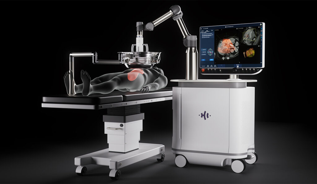
Histotripsy uses high intensity sound waves to “liquify” tumors that have originated in the liver or metastasized to the liver. The non-thermal effects of this focused ultrasound allow for the precise destruction of tissue while minimizing injury to critical structures.
Vanderbilt-Ingram Cancer Center is the only center in a multi-state region to deploy the histotripsy device with the aim of using the procedure to treat liver tumors in adults.
“What’s really exciting about histotripsy is that it allows for mechanical destruction of liver tumors with absolutely no incisions or needles – and no heat – causing minimal damage to surrounding tissue,” said Sekhar Padmanabhan, M.D., an assistant professor of Surgical Oncology and Endocrine Surgery at Vanderbilt University Medical Center. “Recovery time is minimal; patients typically leave the hospital the next day.”
In addition to Padmanabhan, Kamran Idrees, M.D., and Marcus Tan, M.D., will be performing the histotripsy procedure.
“What’s really exciting about histotripsy is that it allows for mechanical destruction of liver tumors with absolutely no incisions or needles – and no heat – causing minimal damage.”
“Besides being completely non-invasive, another key advantage of histotripsy is its ability to preserve important anatomical structures while destroying cancer cells,” said Idrees, division chief of Surgical Oncology and Endocrine Surgery. “It affects the tumor cells, but it doesn’t affect the key structures within the liver such as blood vessels and the bile ducts.”
A significant advantage is its ability to be used repeatedly with patients who develop new tumors over time.
“It’s not one and done,” Idrees said. “If a patient has more lesions or other lesions pop up, we can repeat the procedure as needed.”
Tiny Bubbles to Shockwaves
Histotripsy’s focused ultrasound treatments include the partial or complete destruction of unresectable liver tumors.
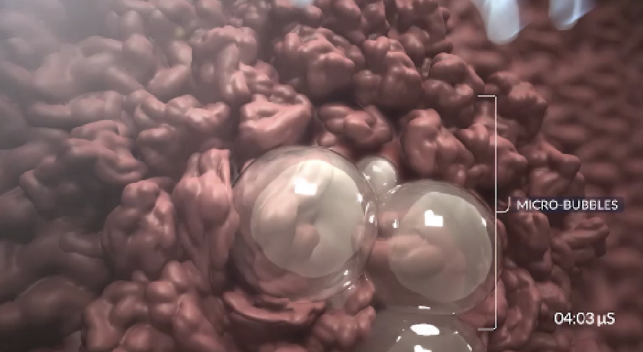
The acoustic waves generate small gas bubbles, or “bubble clouds,” within the targeted tissue. When these bubbles are destroyed, a shockwave is generated that mechanically destroys cell membranes.

“The bubble cloud generated by the short bursts of high frequency ultrasound causes acoustic cavitation, or the generation and oscillation of microbubbles, which causes mechanical strain on cells,” Padmanabhan said.
“This ultimately results in fracturing of cell membranes, destruction of tissue, and release of acellular debris. The technique can be very precise. The bubbles are easy to see with ultrasound, which allows for better targeting.”
In fact, the tumor must be visible on ultrasound for the technology to work, the surgical oncologist explained. The maximum treatment depth is 14 to 16 centimeters, making some tumors difficult to access.
“As for all tumors, we have to make sure we match the right patient and the right tumor – including size and location – with the right treatment.”
Multiple Patient Benefits
Histotripsy is performed under general anesthesia to prevent any movement by the patient while receiving high-intensity ultrasound from outside the body.
“This non-invasive approach makes for a very easy recovery,” Padmanabhan said. “In the study, about 22 percent of participants reported some abdominal pain following the procedure, requiring only Tylenol or Motrin to resolve the pain.”
One particularly notable benefit of histotripsy is that patients can continue their chemotherapy before and after.
“For any surgery and other procedures that we do, we stop chemotherapy for at least four to six weeks,” Idrees said. “And if they have a complication, there may be a delay in restarting the chemotherapy. With histotripsy, we don’t have to worry about any of that.”
Another key benefit is patients can continue blood thinners, since the risk of bleeding is negligible. The treatment can also be combined with other approaches, Idrees added.
“For patients with tumors on both sides of the liver, doctors can use histotripsy to treat one side while surgically removing tumors on the other side. Additionally, histotripsy has been approved for ‘downstaging’ liver tumors — reducing their size to make patients eligible for liver transplantation.”
Future Implications
Padmanabhan cautions that histotripsy will not replace surgery and other treatments for some patients.
“As surgeons, we have a lot of different tools we can use to treat patients, but surgery is still indicated in many cases,” he said. “And we will continue to offer thermal ablation techniques and minimally-invasive liver tumor surgery – we’re one of the few sites to offer that.”
Clinically, histotripsy is also being investigated for the treatment of cardiovascular diseases and other cancers beyond liver tumors.
“The idea of not having to make an incision or insert a needle, reducing post-operative, post-procedure recovery is very exciting, but it’s certainly very early,” Padmanabhan said. “There are clinical trials studying this for kidney tumors, and one is opening soon for pancreas tumors, but it’s important to note that it’s still only approved for liver cancer.”
This early model has been studied in tumors up to 3 cm in diameter, so far.
“It may be applicable to larger tumors, but it just hasn’t been studied in a rigorous fashion,” he added.


